You're about to create your best presentation ever

Fetal Circulation Powerpoint Templates

Create your presentation by reusing a template from our community or transition your PowerPoint deck into a visually compelling Prezi presentation.

fetal circulation

Transcript: baby after the birth: fossa ovalis ligamentum teres ligamentum venosum medical umbilical ligaments superior vesicular artey ligamentum arteriosum In fetal circulation the umbilical cord contains one vein that sends oxygenated blood into a baby's body. There are two arteries that remove the deoxygenated blood. In the circulation of baby after birth, the gas exchange takes place in the lungs. When cutting the umbilical cord, the placental circulation system is switched off and the fetal heart shunts close. how are nutrients absorbed? In fetal circulation waste travels out of the fetus through the umbilical cord, into smaller vessels that lie within the villi of the placenta. Maternal blood fills the spaces that surround the villi waste, and can diffuse through the villi tissues. It also can transfer between the mothers blood and the blood of the vessels of the baby. In circulation of baby after birth waste is removed, digestion of food is eliminated through the anus in the form of feces. fetal circulation how are gases exchanged? In fetal circulation the blood of the umbilical cord moves into smaller blood vessels, which lie within the villi of the placenta. Since the placenta is attached to the mother, the maternal blood can fill the spaces surrounding the villi. Nutrients can diffuse through villi tissues and transfer between the mothers blood and the blood vessels of the baby. In circulation of the baby after birth partially digested food, known as chyme, moves through the small intestines where nutrients are absorbed into the blood stream. how is waste removed? by: Mary Albitz what main structures are involved? fetal structure: foramen ovale umbilical vein intra abdominal part umbilical arteries abdominal ligaments ductus arteriosum how does blood flow through the heart and lungs In fetal circulation, the shunt that bypass the lungs is called the foreman oval. It moves blood from the right atrium of the heart to the left atrium. The ductus arteriosus moves blood from the pulmonary artery to the aorta. Oxygen and nutrients from the mothers blood is transferred across the placenta to the fetus. In Circulation of baby after birth blood enters the heart through two large veins. The inferior and superior vena cava, emptying oxygen poor blood from the body into the right atrium. As the atrium contracts, blood flows from your right atrium into your right atrium into your right ventricle through the open tricuspid valve. Logo

Fetal circulation

Transcript: Fetal circulation There are many differences between regular circulation in an adult and in a fetus. In an adult, arteries carry blood away from the heart and veins carry the blood back to the heart where in a fetus the arteries carry non-oxygenated blood back to the heart and veins carry oxygenated blood away from the heart In an adult or child there is more pressure on the left side of heart but in a fetus the increased pressure is on the right side of the heart. there are two main structural differences. A fetus has a Foramen Ovale and ductus arteriosus to help reroute the blood. For most of it's time in the womb the fetuses lungs are collapsed and not functional. Blood flow to the placenta is provided by a pair of umbilical arteries which arise from the internal iliac arteries and enter the umbilical cord. Blood returns from the placenta in the single umbilical vein, bringing oxygen and nutrients to the developing fetus. Fetal circulation sequence Exchange of gases occurs in the placenta. Oxygenated blood is carried by the umbilical vein towards the fetal heart. The ductus venosus directs part of the blood flow from the umbilical vein away from the fetal liver (filtration of the blood by the liver is unnecessary during the fetal life) and directly to the inferior vena cava. Blood from the ductus venosus enters to the inferior vena cava. Increase levels of oxygenated blood flows into the right atrium. In adults, the increase pressure of the right atrium causes the tricuspid valve to open thus, draining the blood into the right ventricle. However, in fetal circulation most of the blood in the right atrium is directed by the foramen ovale (opening between the two atria) to the left atrium. The blood then flows to the left atrium to the left ventricle going to the aorta. Majority of the blood in the ascending aorta goes to the brain, heart, head and upper body. The portion of the blood that drained into the right ventricle passes to the pulmonary artery. As blood enters the pulmonary artery (carries blood to the lungs), an opening called ductus arteriosus connects the pulmonary artery and the descending aorta. Hence, most of the blood will bypass the non-functioning fetal lungs and will be distributed to the different parts of the body. A small portion of the oxygenated blood that enters the lungs remains there for fetal lung maturity. What is a blue baby A 'blue baby' happens when a baby is born but the foramen ovale or the ductus arteriosus has not closed and the baby is not getting any oxygen because the blood is still bypassing the lungs. Prompt treatment is required and the foramen ovale or ductus arteriosus is surgically closed and blood flow is corrected. When the baby is born it takes a breath and starts regular circulation. It is also known as the fetoplacental circulation, which includes the umbilical cord Another difference is that a fetus does not get oxygen from inhaling it like we do and gas exchange does not happens in the lungs, instead it gets oxygen from the umbilical cored that is attached to the mother and the gas exchange happens in the placenta. A fetal hear beats anywhere from 120 beats to 160 beats per minute Fetal circulation: The transportation of blood containing wastes and nutrients around the body in a fetus (an unborn baby). The umbilical arteries then carry the non-oxygenated blood away from the heart to the placenta for oxygenation.

FETAL CIRCULATION

Transcript: How does the fetal circulatory system work? What if those shunts don't follow the blueprints after birth?... If you remember nothing else.... Normally closes due to increased O2 tension. Acetylcholine, bradykinins, and prostaglandin are the chemical mediators which assist in closure of the ductus arteriosus. Aortic blood is shunted into the pulmonary artery. 2-3 times more common in females than males, etiology is unknown. Most common congenital anomaly associated with maternal rubella infection during early pregnancy Premature infants often have a PDA related to hypoxia and immaturity. First line treatment is to use indomethacin to chemically close the PDA. If that is unsucessful, surgical closure of a PDA is achieved by ligation and division of the ductus arteriosus. *Adaptive fetal circulation. *Higher RBC levels. *Higher hemoglobin; up to and over 20 mg/dl. *Left-Shifted Fetal hemoglobin (HbF) p50 of 19 mm Hg compared to adult's p50 of 27 mm Hg. *Increased cardiac output (200 ml/kg/minute) FETAL CIRCULATION Josh Laack March 8, 2016 REFERENCES Patent Foramen Ovale (PFO) Patent Ductus Arteriosus (PDA) Normally closes due to increased SVR (due to separation from the placenta), increased alveolar and arterial oxygen tension with a resultant decreased PVR (due to lung expansion.) The decreased PVR leads to the decreased right heart pressures. A small, isolated patent foramen ovale essentially has no hemodynamic significance. However, if other defects are present (such as pulmonary stenosis), blood deficient in oxygen will be shunted through the foramen ovale. This hypoxemic blood will go straight into the left ventricle, producing readily identifiable cyanosis. A probe patent foramen ovale is present in up to 25% of people. Usually small enough to be considered clinically insignificant. Patent Foramen Ovale (PFO) Patent Ductus Arteriosus (PDA) PDA is considered more serious than PFO Thanks for your time, and enjoy the rest of your day! Fetal Circulatory Changes In a ....... "Cardiovascular System." Human Embryology Organogenesis. Universities of Fribourg, Lausanne, and Bern (Switzerland)., n.d. Web. 26 Feb. 2016 <http://www.embryology.ch/anglais/pcardio/umstellung02.html>. "Circulatory Changes at Birth." Fetal Circulation. University of California, Berkeley, n.d. Web. 26 Feb. 2016. <https://mcb.berkeley.edu/courses/mcb135e/fetal.html>. Brice, Glenn (2016). Advanced Human Physiology notes. University of Wisconsin La Crosse. Morgan, G.E., Mikhail, M.S. & Murray, M.J. (2013). Cliinical Anesthesiology, Fifth Editon. New York: McGraw-Hill. In the fetus, gas exchange occurs in the placenta. The fetal circulation is "shunt-dependent." The presence of fetal hemoglobin and a high combined ventricular output (CVO) help maintain oxygen delivery in the fetus despite low oxygen partial pressures. The transition from fetal to neonatal life involves closure of circulatory shunts and acute changes in pulmonary and systemic vascular resistance. Overview of Fetal Circulatory Development The system begins to develop toward the end of the third week after conception. The heart starts to beat at the beginning of the fourth week. The most critical period of heart development is from day 20 to day 50 after conception (in a time frame where many women aren't even aware they are pregnant yet...) Many crucial steps occur during cardiac and pulmonary development, and any deviation from this "normal pattern" can cause congenital heart defects, as well as pulmonary complications. 1) Ductus Arteriosus Allows blood to bypass the pulmonary circulation, connects to the descending aorta. Acts to protect the lungs against circulatory overload. Closure starts approximately 10 hours after birth, and is usually complete roughly 3 weeks later. 2) Foramen ovale Shunts highly oxygenated blood from the right atrium to the left atrium. Should close within several hours after birth. 3) Ductus Venosus The fetal blood vessel connecting the umbilical vein to the IVC Carries mostly highly oxygenated blood. Closes mechanically with cord clamping at birth. A stressed newborn can quickly revert back to fetal circulation by reopening the ductus arteriosus and foramen ovale, which can be detrimental to the neonate. Stress factors which anesthesia can help to control include: hypoxemia, hypotension, hypothermia...(3 hypos), as well as hypercapnea and hypervolemia...(2 hypers). Chronic Hypoxia - How Does the Fetus Cope? 3 Shunts in the Fetal Circulation The fetus is connected by the umbilical cord to the placenta, which is the organ that develops and implants in the mother's uterus during pregnancy. Through the blood vessels in the umbilical cord, the fetus receives all the necessary nutrition, oxygen (as fetal lungs do not pacipate in oxygen exchange), and life support from the mother through the placenta. Waste products and carbon dioxide from the fetus are sent back through the umbilical cord and placenta to the mother's circulation to be eliminated.

FETAL CIRCULATION

Transcript: How are wastes removed? FETAL CIRCULATION In fetal circulation the The blood of the umbilical cord moves into smaller blood vessels that lie within the villi of the placenta. Since the placenta is attached to the mother, maternal blood can fill the spaces that surround the villi. Nutrients can diffuse through the villi tissues and transfer between the mother’s blood and the blood vessels of the baby. In circulation of Baby after Birth Partially digested food (chyme) moves through the small intestines where nutrients are absorbed into the bloodstream. In fetal circulation the umbilical cord contains one vein that sends oxygenated blood into baby’s body, and two arteries that remove the deoxygenated blood. In Circulation of Baby after Birth ,The gas exchange takes place in the lungs.By cutting the umbilical cord, the placental circulation system is switched off and The fetal heart shunts become closed. http://mcb.berkeley.edu/courses/mcb135e/fetal.html What main structures are involved? Fetal Structure: -Foramen Ovale -Umbilical Vein (intra-abdominal part) -Ductus Venosus -Umbilical Arteries and abdominal ligaments -Ductus Arteriosum In fetal circulation The shunt that bypass the lungs is called the foramen ovale. It moves blood from the right atrium of the heart to the left atrium. The ductus arteriosus moves blood from the pulmonary artery to the aorta. Oxygen and nutrients from the mother's blood are transferred across the placenta to the fetus. In Circulation of Baby after Birth Blood enters the heart through two large veins, the inferior and superior vena cava, emptying oxygen-poor blood from the body into the right atrium. As the atrium contracts, blood flows from your right atrium into your right ventricle through the open tricuspid valve. -http://www.webmd.com/hypertension-high-blood-pressure/hypertension-working-heart References How does blood flow through the heart and lungs? -http://www.embryology.ch/anglais/pcardio/umstellung02.html -FLVS LESSON -https://www.urmc.rochester.edu/Encyclopedia/Content.aspx?ContentTypeID=90&ContentID=P02362 How are nutrients absorbed? Baby after Birth: -Fossa Ovalis -Ligamentum teres -Ligamentum venosum -Medial umbilical ligaments, superior vesicular artery (supplies bladder) -Ligamentum arteriosum How are gases exchanged? In fetal circulation waste travels out of the fetus through the umbilical cord, to smaller vessels that lie within the villi of the placenta. Maternal blood fills the spaces that surround the villi wastes can diffuse through the villi tissues and transfer between the mother’s blood and the blood vessels of the baby. In circulation of Baby after Birth waste is removed after the digestion of food and is eliminated through the anus in the form of feces.

Fetal Circulation

Transcript: Importance of the Placenta Fetal Circulation Blood Flow Umbilical Arteries Umbilical Cord and Ducutus Venosus Second Shunt Includes large umbilical vein and two smaller umbilical arteries Umbilical vein carries nutrients and oxygen rich blood to the fetus Umbilical arteries carry carbon dioxide and debris laden blood from the fetus to the placenta The ductus venosus bypasses the immature liver and deposit blood through the inferior vena cava which then goes on to the right atrium. At Birth Two Shunts used to bypass lungs Foramen ovale: blood entering right atrium shunted into the left atrium through the opening in the interatrial septum Fetal Tissue A newborn baby has about one cup of blood in circulation The fetal heart rate is approximately twice as fast as an adult's The human placenta is about nine inches long and one inch thick, and weighs around one pound In some cultures the placenta is incinerated, eaten, or buried (cc) image by nuonsolarteam on Flickr Placenta Aorta Collapsed and nonfunctional lungs Foramen ovale closes Ductus arteriosus collapses and turns into fibrous ligamentum arteriosum When blood stops flowing through umbilical vessels they are obliterated Circulatory patterns becomes that of an adult Spark Nutrient, excretory, gas exchanges occur here Lungs and digestive system do not function Nutrients and oxygen move from the mother’s blood into the fetal blood Fetal wastes move in opposite direction Blood that accidently enters right ventricle is pumped out the pulmonary trunk and meets the second shunt: ductus arteriosus A short vessel that connects the aorta and the pulmonary trunk. Blood enters the systemic circulation through shunt Fun Facts!!

Fetal circulation

Transcript: Fetal circulation Simon Androsch What main structures are involved? How does blood flow through the heart and lungs? Fetal Circulation: -Foramen Ovale -Umbilical Vein (intra-abdominal part) -Ductus Venosus -Umbilical Arteries and abdominal ligaments -Ductus Arteriosum Circulation of Baby after Birth: -Fossa Ovalis -Ligamentum teres -Ligamentum venosum -Medial umbilical ligaments, superior vesicular artery (supplies bladder) -Ligamentum arteriosum How are wastes removed? Fetal Circulation:the foraman ovale moves blood through the right atrium of the heart to the left atrium, Then the ductus arteriosus transports blood from the pulmonary artery to the aorta., from there the oxygen and nutrients from the mother's blood are transferred across the placenta to the fetus. Circulation of Baby after Birth: blood enters the right atrium from the large veins where the oxygen poor blood is dumped, when the right atrium then contracts, the blood flows from the right ventricle into the tricuspid valve. How are nutrients absorbed? Fetal Circulation: waste travels out the fetus into the mother by umbilical cord where it is absorbed into the villi in the placenta Circulation of Baby after Birth:the waste of the baby after birth is through the rectum How are gases exchanged? Fetal Circulation: the villi tissues in the mother and are transported through the blood vessels to the baby Circulation of Baby after Birth: chyme travels through the small intestines where the nutrients are absorbed in the blood Fetal Circulation: oxygenated blood enters the babies body from the umbilical cord. and then two arteries remove the deoxygnated blood. Circulation of Baby after Birth: gases are exchanged in the lungs through the alveoli which diffuse carbon dioxide and oxygen

Fetal Circulation

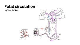
Transcript: Fetal Vs Newborn Circulation By Maria 11/07/22 Introduction Start Fetal development takes place in the uterus. In the uterus, the fetus is surrounded by the amniotic sac and fluid. These features and the underdeveloped state of the organs make it impossible for the fetus to perform respiration and digestion. As a result, both the mother and fetus develop unique structures to allow oxygen and nutrients to make its way into and throughout the fetus's body. Fetal Anatomy The fetus's circulatory system is set up in a way that makes it possible for the fetus to absorb all things necessary for growth and life through the placenta. Fetal circulation completely bypasses the lungs as the fetus can not perform respiration until after birth. The system also bypasses the liver as is also not fully functional until after birth. Fetal anatomy Unique Structures Unique Structures The circulatory system is much more intricate during the fetal stage than after birth. Most of the circulatory, pulmonary, and digestive functions are performed by the mother during this stage of development. Because of this, unique structures such as the placenta, the umbilical arteries, etc develop. They perform many important functions. For example, the placenta and umbilical cord act as a filter for nutrients and oxygen from the mother's blood. They also remove waste from the fetal circulatory system. As a result of the mother providing these necessities and performing these functions, circulatory shunts such as the ductus venosus, the foramen ovale, and the ductus arterious are necessary to the bypassing of the fetal lungs and liver. The fetal liver and lungs are completely unnecessary so, the shunts make for an efficient use of energy and resources. The Placenta Placenta The placenta is a disk-like organ that is attached to the uterine wall during pregnancy and is made of cells contributed by both mother and fetus. The main functions of the placenta are to provide the fetus with oxygen, nutrients, and waste removal through the umbilical cord. Without the placenta to perform these functions, the fetus would not get the nutrients, oxygen, and protection from disease crucial for it's survival and growth. In simple terms, the placenta is necessary because the digestive and pulmonary systems of the fetus are not fully functional and are bypassed by the circulatory system. If the placenta malfunctions, it may result in low birth weight, birth defects, premature birth, and the fetus being more prone to infection. 5-30 minutes after birth, the placenta is also delivered by the mother. If this third stage of labor is not successful, It may lead to hemorrhaging and infection. How does blood flow through the placenta? Blood from the mother fills intervillious space (space between chorionic villi) where nutients and oxygen from the mother are exchanged with deoixygenated blood and waste from the fetus by the maternal pulse. All of this is conducted through the umbilical cord. Fun facts- The placenta also produces hormones to help the fetus grow Ductus Venosus The ductus venosus is one of the three shunts in the fetal circulatory system. This shunt's main purpose is to allow for the passage of oxygenated blood from the umbilical cord through the inferior vena cava and the rest of of the body excluding the liver. This shunt is extremely important because the liver is not fully functional until after the birth of the fetus. Issues with, or a lack of, the ductus venosus can lead to many issues with the fetus such as congenital anomalies (ie. facial clefts, hemivertebrae, cardiac, genitourinary, and gastrointestinal.) After birth, blood pressure in the umbilical sinus decreases and as a result, the shunt retracts, narrows, closes and becomes the ligamentum venosum. Fun fact: Prostaglandin acts to keep the shunt open. Ductus venosus Foramen Ovavle Foramen ovavle Purpose & why: The foramen ovlae is the hole found between the left and right atria of the fetus. The opening's purpose is to to make the bypassing of the pulmonary system possible by allowing blood to pass between the atria. This is important because the lungs are not functional untill after the birth of the fetus and the "shortcut" helps avoid the heart straining itself by pumping blood to unecessary structures(ie. the lungs). Soon after birth when pulmonary circulation is made possible and blood pressure rises in the left side of the heart, tissue grows to close the opening. This results in separation between deoxygenated blood in the right atrium and oxygenated blood in the left atrium. However, if tissue fails to close the hole properly, the probability for strikes is greatly increased as clots can travel from the right to left atrium and later all over the body. Ductus arteriosus Ductus arteriosu Purpose & why: The ductus arteriosus is another of the fetal circulatory shunts. It is a found between the pulmonary artery and the aorta. It sends deoxygenated blood to the lower body of the fetus so that

Now you can make any subject more engaging and memorable